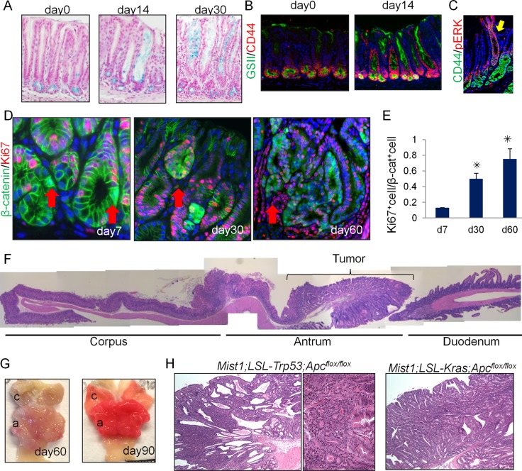
Figure 3. Antral Mist1+ cells serve as a cellular origin of cancer.
(A) Alcian blue (blue) staining in Mist1-CreERT; LSL-KrasG12D mice at the indicated time points. (B) GSII (green) and CD44 (red) staining in Mist1-CreERT; LSL-KrasG12D mice at the indicated time points. (C) CD44 (green) and p-ERK (red) staining in Mist1-CreERT; LSL-KrasG12D mice at day 30 after tamoxifen. (D-E) Immunofluorescence for β-catenin (green) and Ki67 (red) in Mist1-CreERT; Apcflox/flox mice on days 7, 30, and 60 after TAM induction (D). The arrows indicate the nuclear β-catenin+ cells. Ki67+ cell ratio in total nuclear β-catenin+ cells is quantified (E). A total of 300 cells from three mice are analyzed at each time point. (F) Longitudinal H&E stained section of Mist1-CreERT; Apcflox/flox mouse stomach 60 days after TAM induction. (G) Gross picture Mist1-CreERT; Apcflox/flox mice 60 and 90 days after TAM induction. (H) H&E staining of Mist1-CreERT; LSL-Trp53R172H; Apcflox/flox mice and Mist1-CreERT; LSL-KrasG12D; Apcflox/flox mice 150 days after TAM induction.

