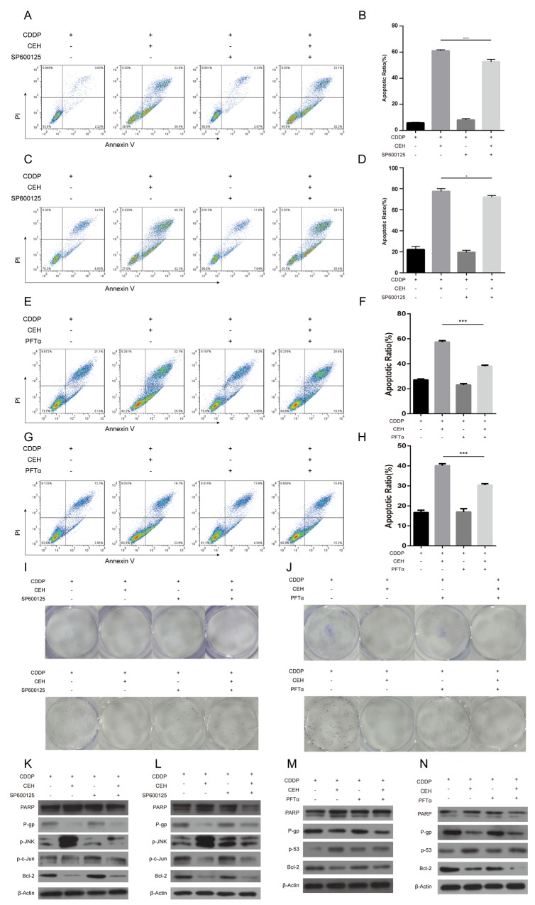
Figure 5. JNK inhibitor SP600125 and p53 inhibitor PFTα can partially reverse the apoptosis and cycle arrest induced by combined cisplatin (cDDP) and cepharanthine hydrochloride (CEH) treatment in Eca109 and Eca109/CDDP cells.
(A-D) Cells were treated with 5 μM CEH and 5 μM SP600125 alone or in combination, in addition to cDDP for 48 h; induction of apoptosis in Eca109 cells (A, B) and Eca109/CDDP (C, D) evaluated by Annexin-V-FITC/PI staining. (E-H) Cells were treated with 5 μM CEH and 0.5 μM PFTα alone or in combination, in addition to cDDP for 48 h; induction of apoptosis in Eca109 (E, F) and Eca109/CDDP (G, H) cells evaluated by Annexin-V-FITC/PI staining. (I and J) Cells were treated with 5 μM CEH, 5 μM SP600125 (I) or 0.5 μM PFTα (J) alone or in combination, in addition to cDDP for 48 h; representative images of Eca109 (upper panel) and Eca109/CDDP (lower panel) clone formation are shown. (K-N) Western blotting detection of PARP, P-gp, phosphorylated JNK (p-JNK), phosphorylated c-Jun (p-c-Jun) and Bcl-2 protein expression in Eca109 (K, M) and Eca109/CDDP (L, N) cells treated with 5 μM CEH, 5 μM SP600125 or 0.5 μM PFTα alone or in combination, in addition to cDDP for 48 h. β-Actin was used as an internal control. Data represent the mean ± SD of at least three independent experiments. Data are presented as mean ± SD. *, P < 0.05; **, P < 0.01; ***, P < 0.001.

