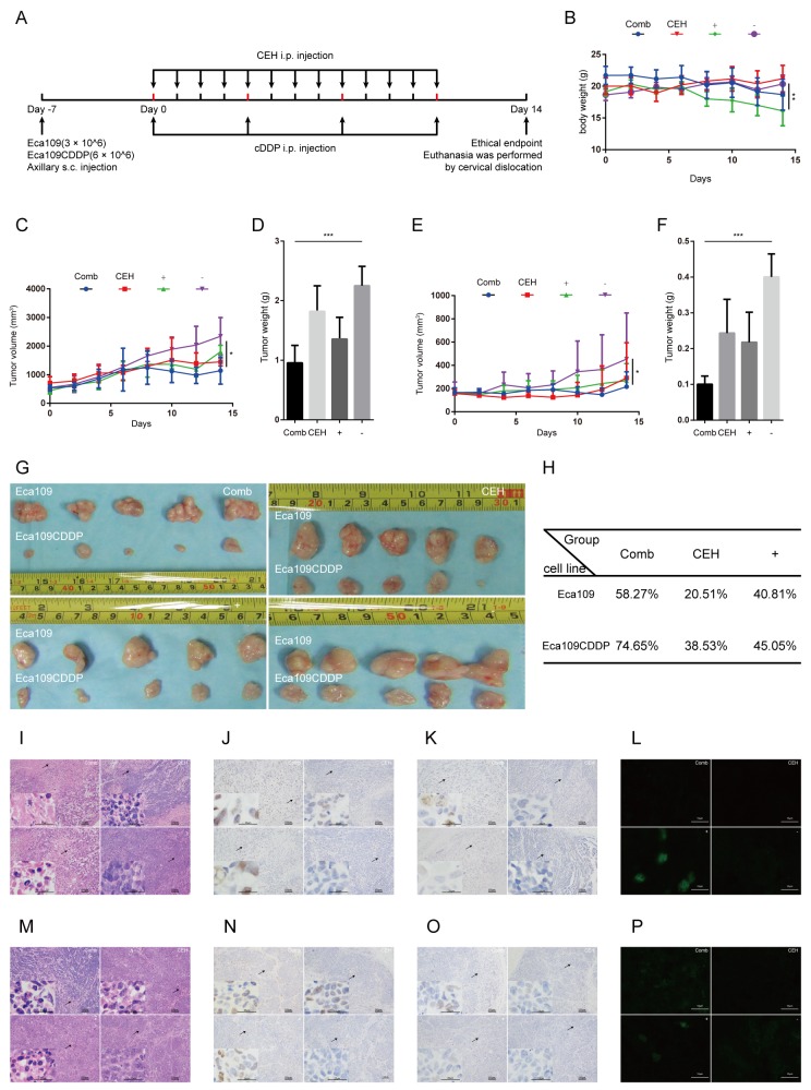
Figure 6. Cepharanthine hydrochloride (CEH) combined with cisplatin (cDDP) inhibits esophageal squamous cell carcinoma xenograft tumor growth.
(A) Time line of esophageal squamous cell carcinoma cell inoculation and drug treatment. (B) Time courses of animal weight. (C) Time courses of Eca109 cells xenograft tumor growth. (D) Bar graphs represent the mean weight of the Eca109 cells xenograft tumor after various treatments. (E) Time courses of Eca109/CDDP cells xenograft tumor growth were measured in each group at the indicated time point of various treatments. (F) Bar graphs represent the mean weight of the Eca109/CDDP xenograft tumor after various treatments. (G) Visual comparison of the dissected tumor tissues. Representative pictures of tumor growth in mice treated with vehicle control and the various treatments are shown. (H) Effects of CEH combined with cDDP on tumor inhibition rate of tumor mice. (I and M) Images are the representative H&E stained esophageal squamous cell carcinoma xenografts, originating from Eca109 cells (I) and Eca109/CDDP cells (M). (J and N). Expression of c-Jun in tumor tissues, originating from Eca109 cells (J) and Eca109/CDDP cells (N), was assessed by immunostaining, scale bars: 25 μm. (K and O) Expression of phosphorylated c-Jun in tumor tissues, originating from Eca109 cells (K) and Eca109/CDDP cells (O), was assessed by immunostaining, scale bars: 25 μm. (L and P) Expression of P-gp in tumor tissues, originating from Eca109 cells (L) and Eca109/CDDP cells (P), was assessed by immunofluorescence, scale bars: 25 μm. Data are presented as mean ± SD. **, P < 0.01; ***, P < 0.001. N = 5 in each group.

