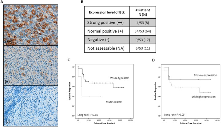
Figure 4. Btk IHC expression in HRS cells and Its correlation with survival.
(A) Representative examples of IHC for Btk expression in cHL tissues. (B) Distribution of Btk protein expression in our series. Positivity (+) was concluded for cases with a level of expression comparable to that seen in normal germinal center B lymphocytes. (C) Kaplan–Meier survival curves demonstrate longer FFS in wt-BTK cases (P<0.05). (D) Survival curves demonstrate a longer FFS in cases with a low level of expression of Btk protein (P=n.s.).

