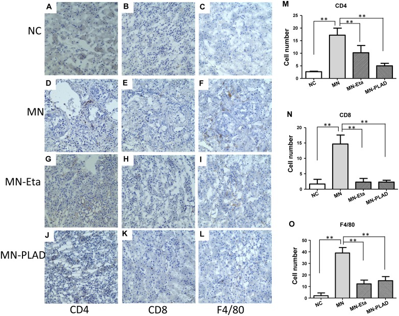
Figure 7. Immunohistochemistry for CD4, CD8, CD19, and F4/80 in kidneys.
Kidneys from mice in the NC group (A–C) and MN group (D–L), which were treated with PBS (D-F), etanercept (MN-Eta; G-I), or preligand assembly domain fusion protein (MN-PLAD; J-L) were stained for anti-CD4 (A, D, G, J), anti-CD8 (B, E, H, K), and anti-F4/80 (C, F, I, L). Quantitative data are shown in (M-O). All images are at 400× magnification. *p < 0.05 versus the control group.

