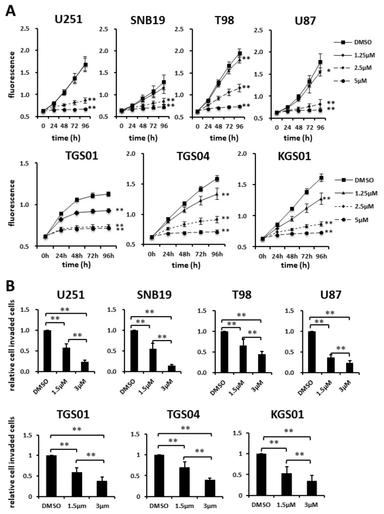
Figure 3. Effects of fluspirilene on proliferation and invasion of GBM cells.
(A) Cells were treated with fluspirilene at 1.25, 2.5, and 5 μM, and viability curves of GBM cell lines were analyzed in the presence and absence of fluspirilene. The plate was read on a plate reader at the indicated time points. (B and C) Cells were treated with fluspirilene at 1.5 and 3 μM. Cells invading through a Matrigel-coated Transwell chamber were scored in the presence and absence of fluspirilene for 8 h. The mean numbers of cells and standard deviations were calculated for eight high-power microscopic fields. *p < 0.05, **p < 0.01.

