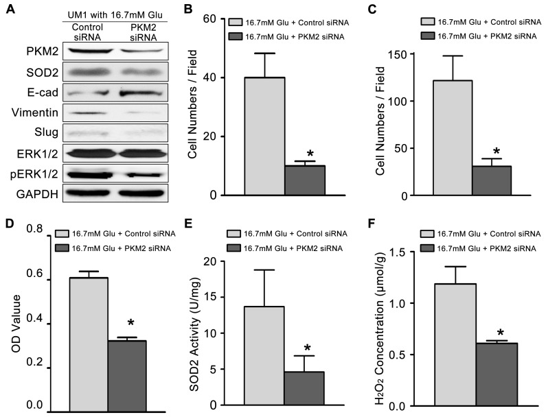
Figure 3. High glucose mediated the migration and invasiveness of TSCC through PKM2 pathway.
(A) The protein levels of PKM2, SOD2, Vimentin, Slug and pERK1/2 were decreased in UM1 cells after knockdown of PKM2 in TSCC cells cultured with 16.7 mM glucose, but the protein level of E-cadherin was decreased. PKM2 knockdown inhibited the migration (B) and invasion (C) abilities, cell proliferation (D), and the SOD2 activity (E) and H2O2 production (F) in UM1 cells cultured with 16.7 mM glucose. *P<0.05 compared with UM1 cells cultured with 16.7mM glucose and transfected with control siRNA.

