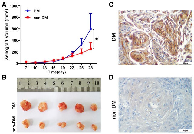
Figure 4. High glucose promotes tumour growth of TSCC in vivo.
(A-B) CAL27 cells were inoculated subcutaneously into BALC/C nude mice model of DM. Tumour growth was significantly slower in the non-DM group relative to the DM group. (C-D) PKM2 expression was obviously increased in TSCC xenografts from DM group compared with the non-DM group detected by IHC. Scale bar: 50 μm.

