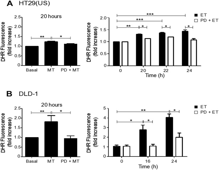
Figure 6. MEK inhibition reduces ROS production induced by Methotrexate and Etoposide.
(A) HT29(US) or (B) DLD-1 colon cancer cells (3 x 10⁶) were seeded in 24-well plates and, after 24 h, were treated with the MEK inhibitor PD98059 (PD, 50 μM) for 30 min, added prior to treatment with 100 nM Methotrexate for 20 h or 10 μM Etoposide for the indicated time periods. Cells were washed 3 times with PBS and subsequently incubated with trypsin for 5 min. Once in suspension, cells were loaded with the probe DHR123 (1.4 μg/ml) in RPMI media without serum for 30 min and then the reaction was stopped on ice. The extent of DHR123 oxidation was determined by flow cytometry. The graphs show DHR123 fluorescence normalized to the untreated condition (Basal) (mean ± SEM) averaged from 3 independent experiments. Significant differences are indicated ***p≤ 0.001, **p ≤ 0.01, *p ≤ 0.05.

