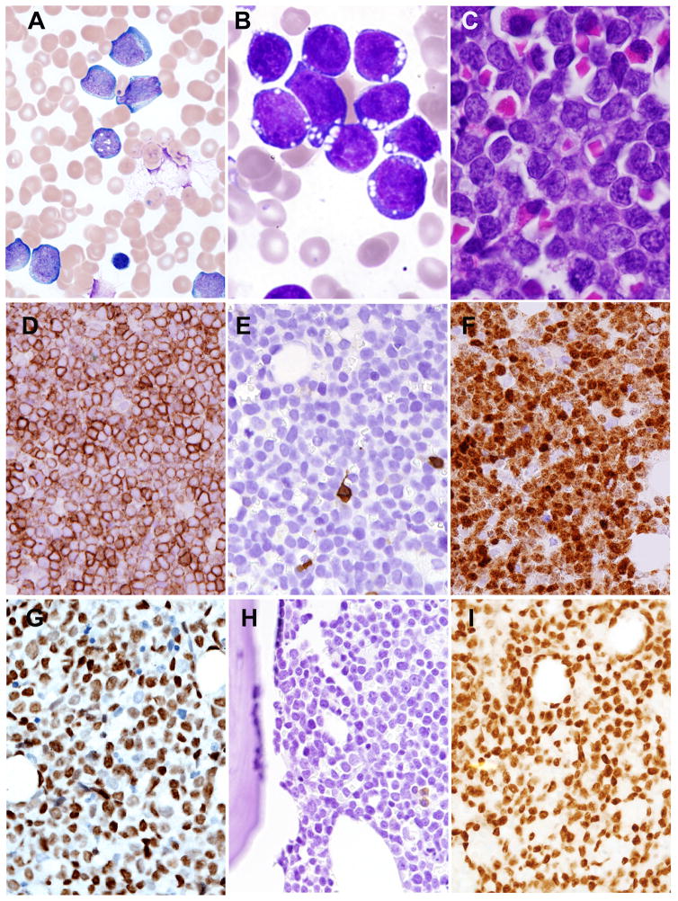
Fig. 1. Bone marrow and peripheral blood findings of MYC+ B-ALL/LBL.
A: Blasts in peripheral blood smear (Wright-Giemsa stain, 1000× (case 2) were intermediate-in-size, with ovoid-to-irregular nuclei, fine chromatin, occasional prominent nucleoli, and scant-to-moderate deeply basophilic cytoplasm. B: Bone marrow aspirate smear, case 1 (Wright-Giemsa stain, 1000×) showed cells with high nuclear:cytoplasmic ratio, deeply basophilic cytoplasm, and cytoplasmic vacuoles. C: Bone marrow core biopsy (H&E 1000×, (case 1) showed a diffuse infiltrate of blasts with finely dispersed chromatin and small nucleoli. Immunohistochemical findings (Case 1) showed positive staining for CD19 (D), but absence of CD20 (E). TDT showed strong nuclear staining (F). The blasts show nuclear staining for MYC (Case 2) (G). BCL-6 was negative (H), but PAX5 was positive (I).

