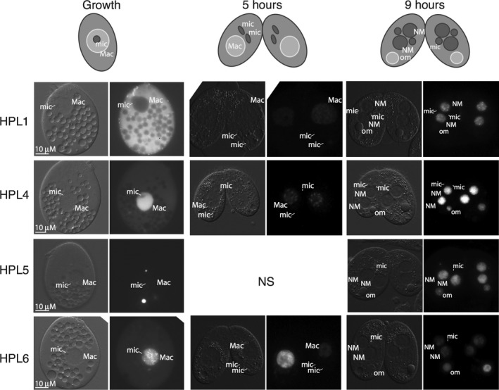
Figure 3.

Hpl proteins localize to macronuclear chromatin, new developing macronuclei, and pycnotic nuclei. YFP fusion proteins were expressed by induction with CdCl2 and visualized by epifluorescence microscopy during regular growth, 5 h into conjugation, and 9 h into conjugation. Schematic at the top shows arrangements of various nuclei. DIC images on the left of each image pair show positions of nuclei. NS = not shown; NM = new macronucleus; mic = micronucleus; Mac = macronucleus; om = old macronucleus. Scale bars = 10 μm apply to all panels in the set.
