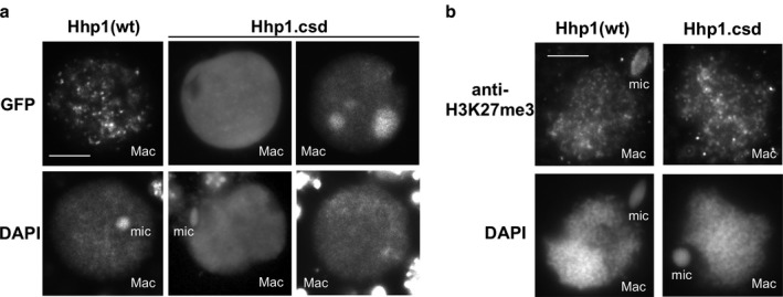
Figure 5.

Loss of the CSD reduces chromatin body targeting of Hhp1 in nuclei from growing cells. (a) Cells were induced to express GFP‐Hhp1Δcsd (“Hhp1.csd”), then stained with DAPI and visualized by epifluorescence microscopy. Two sets of nuclear images representing two observed localization patterns of GFP‐Hhp1Δcsd are shown. Scale bar = 5 μm shown in first panel applies to all images. (b) Cells expressing GFP‐Hhp1Δcsd were subjected to immunofluorescence with anti‐H3K27me3, counterstained with DAPI, and visualized by epifluorescence microscopy. Mac = macronucleus; mic = micronucleus. Scale bar = 5 μm shown in first panel applies to all images.
