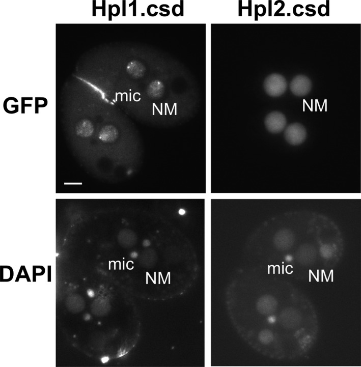
Figure 6.

Loss of the CSD affects localization of Hpl2, but not Hpl1. Cells were induced to express GFP‐Hpl1Δcsd (“Hpl1.csd”) or GFP‐Hpl2Δcsd, then stained with DAPI and visualized by epifluorescence microscopy. NM = new macronucleus; mic = micronucleus. Scale bar = 10 μm shown in first panel applies to all.
