. 2017 Dec 26;30(1):47–53. doi: 10.5021/ad.2018.30.1.47

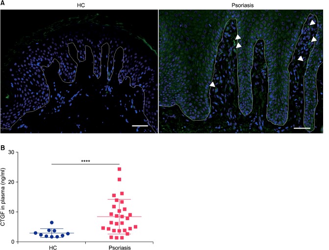
Fig. 4. Connective tissue growth factor (CTGF) expression was increased in skin lesions and plasma from psoriasis patients. (A) Representative immunofluorescent staining of CTGF (green) in skins of healthy control (n=4) and psoriasis patients (n=6). Nuclei were stained with 4′,6-diamidino-2-phenylindole (blue). Arrowheads showed CTGF-expressing cells; bar: 50 µm. White line represents the border between the epidermis and dermis. (B) CTGF in plasma from healthy control (HC) and psoriasis patients was detected by a sandwich enzyme-linked immunosorbent assay (ELISA) analysis. Blue circle: HC (n=10), red rectangle: psoriasis patients (psoriasis, n=28). Values are shown as the mean±standard deviation of the mean. ****p<0.0001 by unpaired t-test with Welch's correction.

Fig. 4