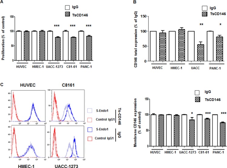
Figure 5. Effect of TsCD146 mAb antibody on cell proliferation and CD146 expression.
(A) Analysis of the proliferation of HUVEC, HMEC-1, UACC-1273, C81-61 and Panc-1 cells after 72 h in the presence of IgG or TsCD146 mAb (5 μg/ml). The results are expressed as mean values +/− standard deviation of 3 independent experiments. (B) Total CD146 expression was determined by ELISA on lysates of UACC, Panc-1, HUVEC and HMEC-1 cells after 72 h of treatment with control IgG or TsCD146 mAb. Mean values of three independent experiments are shown. (C) Membrane expression of CD146 was determined by flow cytometry with S-endo1 antibody in the different cell lines after treatment with either TsCD146 mAb (5 μg/ml) or IgG (5 μg/ml) for 72 hours. Representative FACS profiles are shown. The results are expressed as mean values +/− standard deviation of 3 experiments. *p < 0.05, **p < 0.01, ***p < 0.001, experimental versus control.

