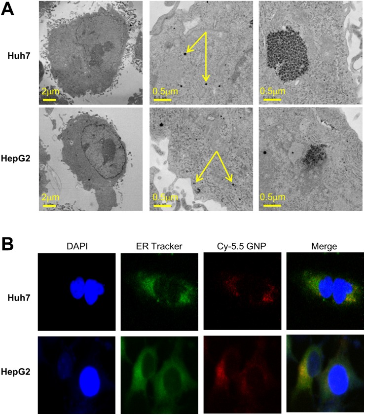
Figure 1. Intracellular localization of 5 nm gold particles.
(A) Transmission electron microscopy (80kV) of monodispersed 5 nm gold nanoparticles. (B) Huh7 and HepG2 cells incubated for 24 h with 1 mM Cy5.5-labeled gold nanoparticles, and stained with specific dyes for nuclei (DAPI) and endoplasmic reticulum.

