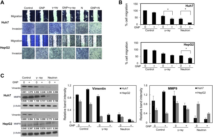
Figure 5. Effect of gold nanoparticles and radiation on cell migration and invasiveness.
(A, B) Plates from the scratch assay were photographed, distances between migrating cell fronts were measured, and the fraction of cells that had migrated was calculated (upper). Values are mean ± SD of three experiments. Cells exposed to γ-rays and neutron radiation (5 Gy, 5 GyE). Cell invasion was examined by Matrigel transwell chamber assay (lower). (C) Immunoblotting of cell lysates with indicated antibodies. Cells exposed to γ-rays and neutron radiation (5 Gy, 5 GyE). Band intensities for target proteins were normalized to that for β-actin. Values represent the means of 3 experiments ± SD. (D) Immunocytochemistry staining for Vimentin in Huh7 and HepG2 cells exposed to γ-rays and neutron radiation (5 Gy, 5 GyE) in the absence or presence of gold nanoparticles. (E) 3D spheroid growth assay of Huh7 and HepG2 cells treated with gold nanoparticles and radiation for four days. Phase-contrast images indicated that untreated cells formed polarized spheroids, but cells exposed to gold nanoparticles and radiation did not. Cells exposed to γ-rays and neutron radiation (5 Gy, 5 GyE).


