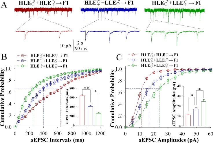
Figure 5. Excitatory synaptic transmission on barrel cortical GABAergic neurons decreases after pairing WS and OS, especially in F1 mice with the high efficiency of odorant-induced whisker motion from HLE parents.
Spontaneous excitatory postsynaptic currents (sEPSC) were recorded on the GFP-labeled GABAergic neurons in cortical slices under voltage-clamp (holding potential at -65 mV) in presence of 10 μM bicuculline. (A) shows sEPSCs recorded on the neurons in CR-formation F1 mice from cross-matings of HLE mice (HLE♂+HLE♀, red traces), HLE female mice and LLE male mice (HLE♀+LLE♂, blue) as well as HLE male mice and LLE female mice (HLE♂+LLE♀, green).. Bottom traces are the expanded waveforms selected from top traces. Calibration bars are 10 pA, 2 second (top) and 90 ms (bottom). (B) shows cumulative probability versus sEPSC intervals in the neurons from CR-formation F1 mice from cross-matings of HLE mice (HLE♂+HLE♀, red symbols), HLE female mice and LLE male mice (HLE♀+LLE♂, blue) as well as HLE male mice and LLE female mice (HLE♂+LLE♀, green). Inserted figure shows the comparisons of sEPSC intervals at 67% cumulative probability from three groups of mice (n = 15 neurons from 8 mice for each group). (C) shows cumulative probability versus sEPSC amplitudes in the neurons from CR-formation F1 mice from cross-matings of HLE mice (HLE♂+HLE♀, red symbols), HLE female mice and LLE male mice (HLE♀+LLE♂, blue) as well as HLE male mice and LLE female mice (HLE♂+LLE♀, green). Insert figure denotes the comparisons of sIPSC amplitudes at 67% cumulative probability from three groups of mice (n = 15 neurons from 8 mice for each group). An asterisk shows p < 0.05, two asterisks show p < 0.01 and three asterisks show p < 0.001, in which the statistical comparisons are one-way ANOVA.

