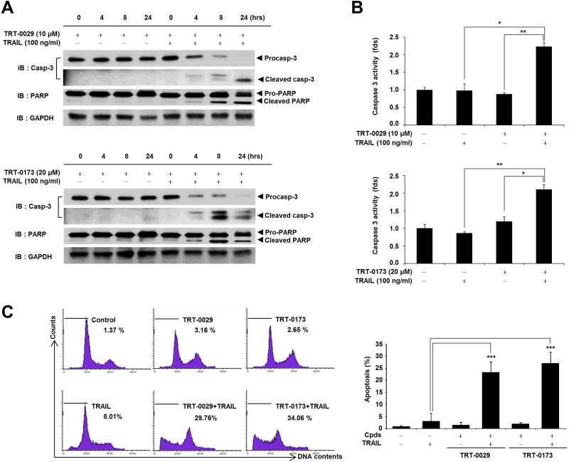
Figure 4. Lead compounds induce TRAIL-mediated apoptosis via caspase signals in Huh7 cells.
(A) Huh7 cells were treated with lead compound (10 μM for TRT-0029 and 20 μM for TRT-0173) in the presence or absence of 100 ng/ml TRAIL for the indicated time periods. The PARP and caspase-3 levels were examined by Western blotting. GAPDH was used as a loading control. (B) Huh7 cells were treated with lead compound (10 μM for TRT-0029 and 20 μM for TRT-0173) and TRAIL for 24 h. The cells were analyzed for caspase-3 activity and differences in this activity were assessed using Student's t-test (*p < 0.05, **p < 0.01) (C) Cell cycle analysis of propidium iodide-stained Huh7 cells was performed using flow cytometry (left). The bars indicate the percentages of cells in the sub-G1 phase based on apoptosis signals. Bar graphs represent the percentages of sub-G1 DNA contents undergoing apoptosis (***p < 0.001) (right).

