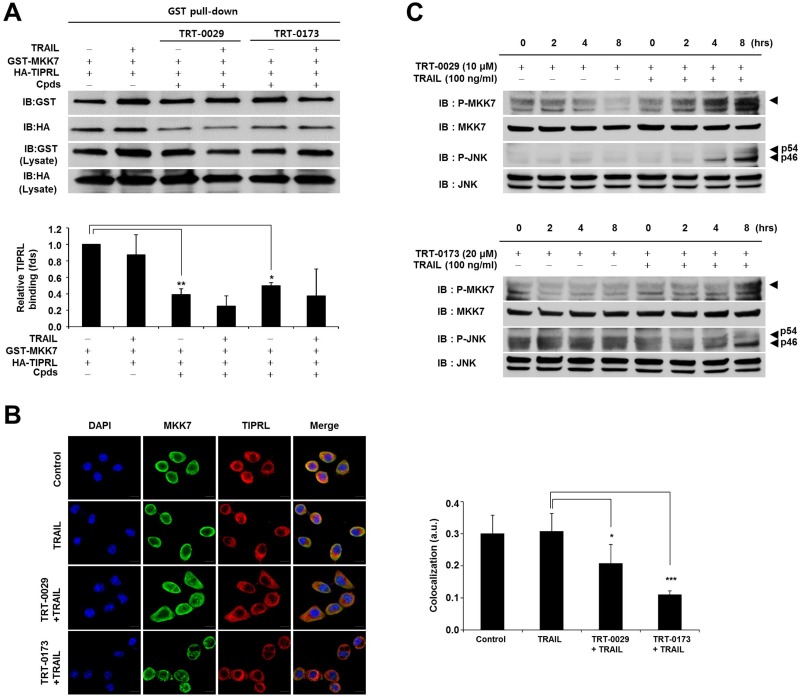
Figure 5. Effects of inhibition of MKK7-TIPRL interaction and activation of MKK7/JNK by the lead compounds.
(A) GST pull-down assay. 293T cells were co-transfected with GST-MKK7 and HA-TIPRL vectors for 24 h and then treated with lead compound (10 μM for TRT-0029 and 20 μM for TRT-0173) and/or TRAIL for 24 h (upper). The signal intensity corresponding to HA-TIPRL/GST-MKK7 was quantified by the ImageJ program, and differences in this activity were assessed using Student's t-test (*p < 0.05, **p < 0.01) (lower) (B) Huh7 cells were treated with lead compound (10 μM for TRT-0029 and 20 μM for TRT-0173) in the presence of TRAIL for 8 h, were stained with antibodies against MKK7 (green) and TIPRL (red), and then observed by confocal microscopy. DAPI (blue) was used for nuclear staining. Scale bar, 50 μm (left). The significance of the differences was assessed with a one-way ANOVA and Bonferroni's multiple comparison post hoc test (*p < 0.05, ***p < 0.001). Yellow indicates the co-localization of TIPRL (red) and MKK7 (green) (right). a.u., artificial unit. (C) Huh7 cells were treated with lead compound in the presence or absence of 100 ng/ml TRAIL for the indicated time periods. The phosphorylation of MKK7 and JNK was examined by Western blotting.

