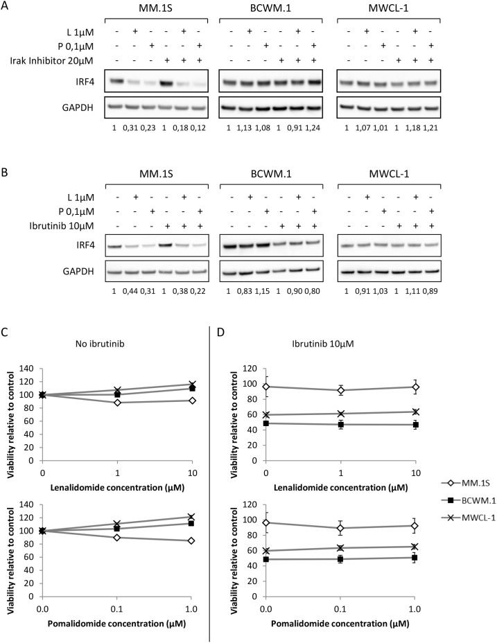
Figure 6. Inhibition of MYD88 and Btk signaling pathways does not sensitize WM cells to lenalidomide and pomalidomide.
(A and B) Western blot analysis using anti-IRF4 mouse monoclonal antibody showing IRF4 expression after 48 hours exposure to lenalidomide and pomalidomide at the indicated concentrations with or without Irak inhibitor (A, 20μM) or Ibrutinib (B, 10μM). Band intensities were quantified using ImageQuant TL software, normalized to their respective GAPDH bands and expressed comparatively to the untreated control. Picture is representative of at least three different experiments. (C and D) Viability (MTS) analysis of the MM or WM cell lines after 48 hours exposure to lenalidomide and pomalidomide at the indicated concentrations, with (C) or without (D) 10μM of Ibrutinib. L: Lenalidomide; P: Pomalidomide.

