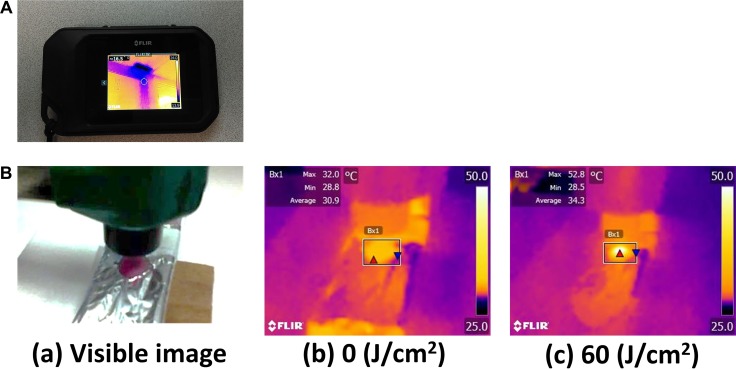
Figure 1. Overview of the thermal imaging camera and thermal image.
(A) The thermal imaging camera used in this study. (B) A visible image and the thermal images taken by the camera before and after NIR irradiation. The maximum temperature exposed to NIR light, was measured. The temperature of mouse skin increased with NIR light irradiation gradually.

