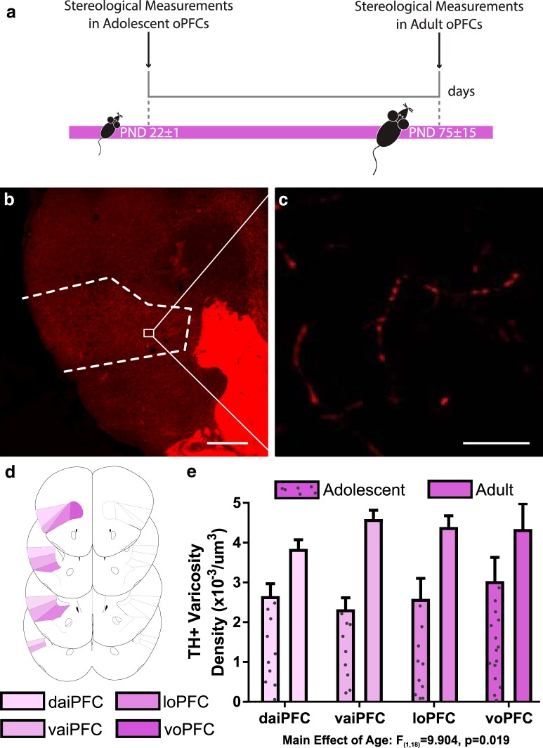
Figure 1.
Dopamine varicosity density in the oPFC is protracted across adolescence. A, Timeline of experimental procedures; n = 4 per group. B, A micrograph of a coronal section through the frontal cortex of an adult mouse at low magnification (4×) showing the contour of the oPFC. Scale bar = 500 μm. C, A micrograph of a coronal section of the oPFC of an adult mouse at high magnification (60×) showing TH-immunopositive varicosities. Scale bar = 10 μm. D, The voPFC, loPFC, vaiPFC, and daiPFC respectively, are highlighted in increasingly pale shades of purple. Line drawings were derived from Paxinos and Franklin (2013). E, Stereological quantification of dopamine varicosity density reveals that there are more dopamine varicosities in the adult oPFC than the adolescent. Bars represent mean ± standard error (Extended Data >Fig. 1-1).

