Figure 2.
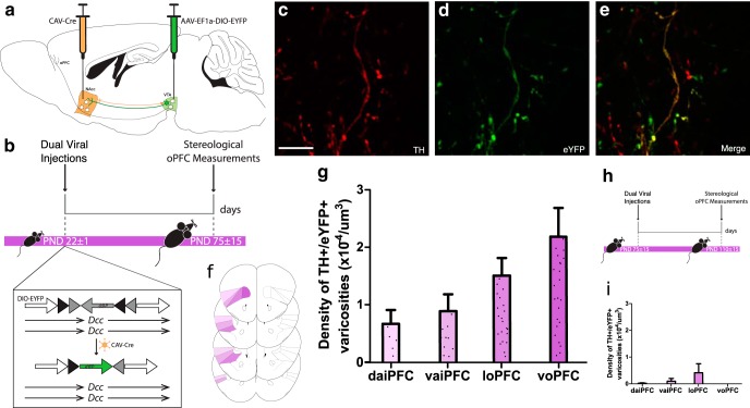
Axons continue to grow to the oPFC during adolescence. A, The dual-viral injection method used to label NAcc-projecting ventral tegmental area neurons with eYFP. B, Timeline of experimental procedures: mice were injected at the start of adolescence (PND22 ± 1) and six weeks later, at which point adolescent mice have reached adulthood, eYFP-expressing dopamine axons in the oPFC were quantified; n = 5. C–E, Micrographs of a coronal section of the oPFC of an adult mouse at high magnification (60×) show (C) TH-immunopositive varicosities, (D) eYFP-expressing varicosities, and (E) an overlay highlighting co-labeled varicosities. Scale bar = 10 μm. F, The voPFC, loPFC, vaiPFC, and daiPFC, respectively, are highlighted in increasingly pale shades of purple. Line drawings were derived from Paxinos and Franklin (2013). G, Stereological quantification of dopamine varicosity density reveals eYFP-expressing dopamine varicosities are present in the oPFC in adult mice that received dual-viral injections in early adolescence (Extended Data Fig. 2-1). H, To ensure the eYFP-expressing dopamine neurons in the oPFC were the result of axon growth and not collaterals, we injected viruses into early adult mice and quantified eYFP-expressing dopamine axons six weeks later; n = 3. I, eYFP-expressing dopamine varicosities are almost entirely absent from the orbital prefrontal cortices of mice that were injected during adulthood (Extended Data Fig. 2-1). G & I, Bars represent mean ± standard error.

