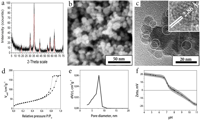
Figure 1.
XRD diffraction pattern of the material corresponds to magnetite oxide phase, peaks referred to JCPDS file No. 19–0629: (red lines) (a); SEM image shows porous microstructure of the material (b); according to TEM, a magnetite matrix consists of truncated tetrahedron nanoparticles (highlighted by white contour) with narrow size distribution (c); low-temperature N2 physisorption curve shows mesoporous structure of the material (d); pore size distribution of the magnetite xerogel matrix by the BJH method (e); zeta potential of the magnetite matrix at different pH values. At pH = 7.4 the matrix is positively charged (f).

