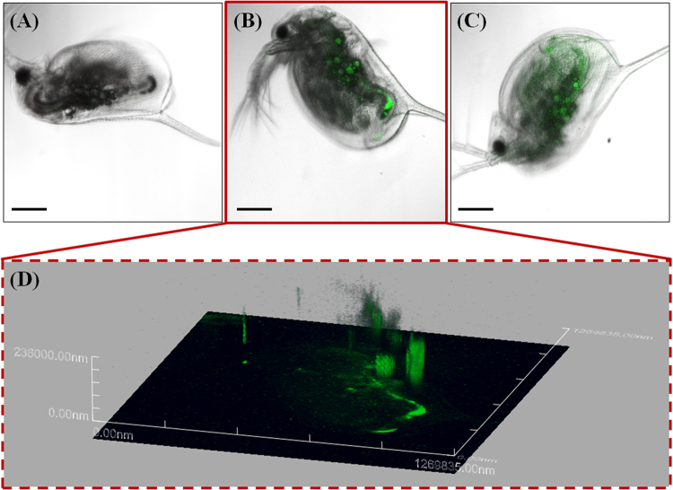
Figure 3.
Confocal laser scanning microscope (CLSM) images of Daphnia magna that were not exposed to nano-sized polystyrene (nPS; green emissions) (a) and that were exposed through the diet (b,c). Z-stack image of individual exposed through the diet (b), observed using CLSM (d). Z-stack image (d) provides evidence of nPS uptake through dietary exposure of D. magna. Green fluorescent nPS was observed in the gut of exposed D. magna. Scale bar = 200 μm.

