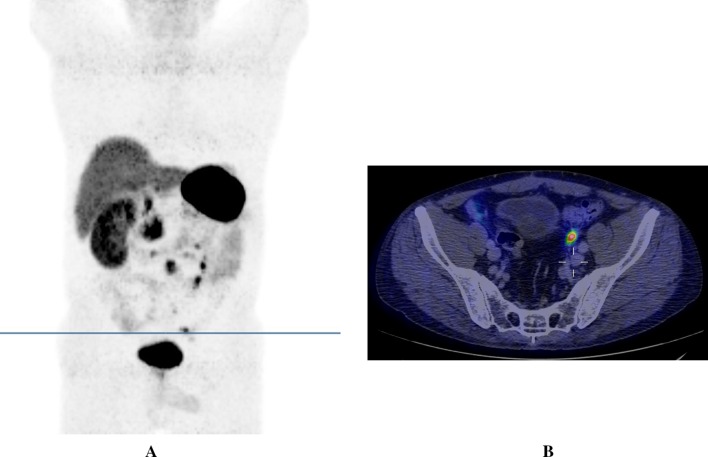
Fig. 1.
68Ga-DOTATOC-PET/CT. 3D PET reconstruction (maximum intensity projection) showing multiple peritoneal carcinomatosis lesions predominately in the left lower part of the abdomen (a). The transverse line indicates a peritoneal metastasis in the left pelvis and corresponds to the level of the transverse PET/CT fusion image (b) in which the same lesion is shown with a high 68Ga-DOTATOC uptake (above the upright post of the cross)

