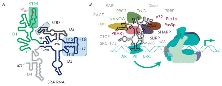
Fig. 6.
Schematic representation of the secondary structure of human SRA RNA (A) and its currently known protein partners (B) according to Liu et al. [61]. SRA RNA domains are colored: D1 – green, D2 – black, D3 – blue, D4 – grey. The U207 residue subjected to pseudouridilation is marked by an asterisk. Panel A shows the main structural elements of SRA RNA that bind several proteins, shown in color frames. Panel B shows a schematic representation of the proteins which directly bind to SRA RNA (they are labeled in corresponding colors). Nuclear receptors (AR – androgen, PR – progesterone, ERα – estrogen α) are colored in blue. All other proteins known to interact with SRA RNA are denoted in grey (without animation).

