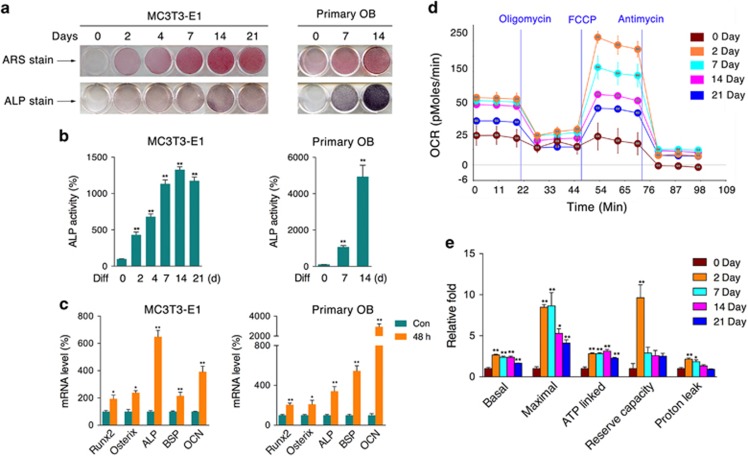
Figure 1.
Increased mitochondrial oxygen consumption in osteoblast differentiation. MC3T3-E1 cells and calvaria-derived primary osteoblasts were induced to differentiate for the indicated periods of time. (a) ARS and ALP staining of differentiated cells. (b) ALP activity of cell homogenates. (c) After differentiation for 48 h, the relative mRNA levels of Runx2, Osterix, ALP, BSP, and OCN were determined by qRT-PCR. The oxygen consumption rate (OCR) of MC3T3-E1 cells at various time points was measured with an XF24 Extracellular Flux Analyzer (d) experiment program and (e) statistical analysis). Data are presented as the mean±S.E.M. from at least three independent experiments. *P<0.05, **P<0.01 versus relative control

