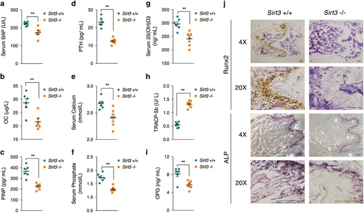
Figure 7.
Serum and histochemical analyses of osteoblast function in Sirt3−/− mice. Serum was collected from wild-type and Sirt3−/− mice, and the levels of the following factors were analyzed: (a) bone alkaline phosphatase (BAP); (b) osteocalcin (OC); (c) procollagen type I N-terminal propeptide (P1NP); (d) parathyroid hormone (PTH); (e) calcium; (f) phosphate; (g) 25(OH)D3; (h) tartrate-resistant acid phosphatase 5b (TRACP-5b); and (i) osteoprotegerin (OPG). (j) Femur sections were prepared from wild-type and Sirt3−/− mice at 8 weeks and immunostained with an anti-Runx2 antibody and an ALP staining kit. Data are presented as the means±S.E.M.; n=6 per group. **P<0.01 versus relative control

