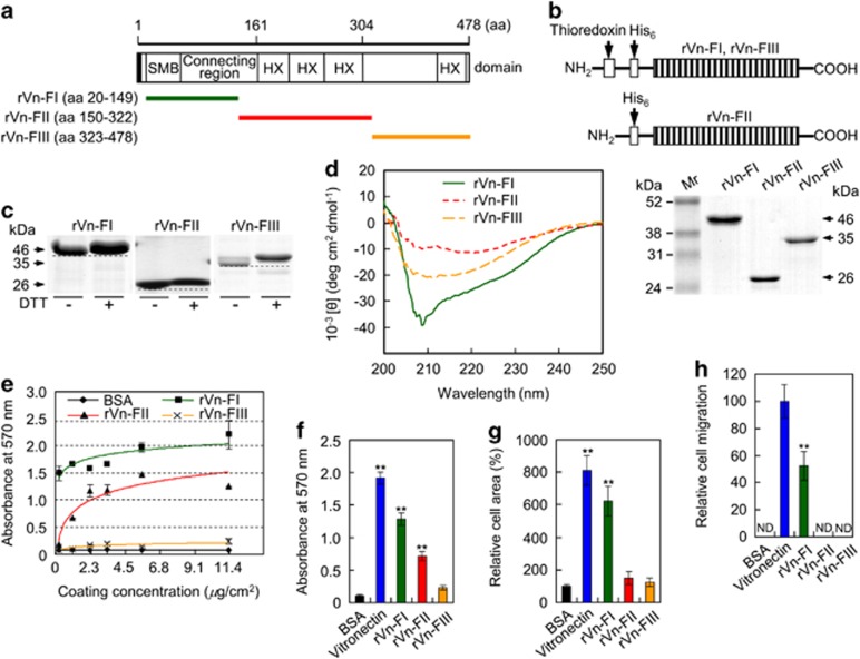
Figure 1.
Analyses of purified rVn truncations by SDS-PAGE and circular dichroism spectroscopy and their effects on cell behavior. (a) Schematic diagram of the rVn truncations. The amino acid (aa) positions and the domain structure of full-length vitronectin are indicated. The black box and colored bars represent the signal peptide and the positions of the recombinant proteins, respectively. (b) Schematic diagram and SDS-PAGE analyses (10% polyacrylamide gels under reducing conditions) of the purified rVn proteins, expressed as His6-tagged fusions. The bands were visualized by Coomassie staining. (c) Gel mobilities (12.5% SDS-PAGE) of the purified rVn proteins treated with or without dithiothreitol (DTT). (d) Circular dichroism analysis of the rVn fragments in PBS (pH 3.0) at 23 °C. (e) The dose-dependent effects of the truncated rVn proteins on the attachment of human osteogenic cells. The cells were seeded onto rVn-treated plates for 1 h in serum-free medium. (f and g) Cell attachment (f) and spreading (g) of osteogenic cells induced by BSA (1%), vitronectin (0.23 μg/cm2), and the truncated rVn proteins (5.7 μg/cm2) for 1 h (f) or 3 h (g) in serum-free medium. (h) Migration of osteogenic cells induced by vitronectin and the truncated rVn proteins. Osteogenic cells were seeded into the upper chambers of Transwell filters coated with vitronectin or rVn proteins and were incubated for 24 h. ND, not detected. Data in (e–h) represent the mean±SD (n=4). **P<0.01

