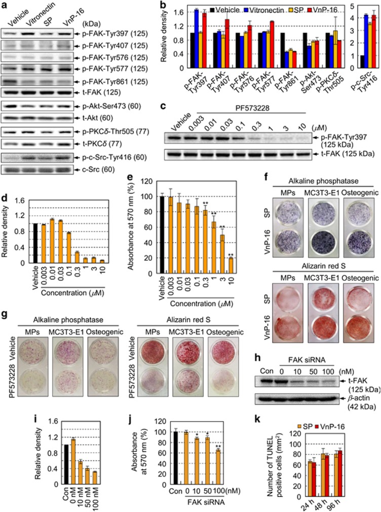
Figure 4.
VnP-16 promotes osteogenic differentiation through β1 integrin/FAK signaling. (a,b) Immunoblotting (a) and densitometric analyses (b) of phospho-FAK, phospho-Akt Ser473, phospho-PKCδ Thr505, and phospho-c-Src Tyr416 in osteogenic cells that were cultured for 3 h on plates coated with vitronectin (0.23 μg/cm2), SP, or VnP-16 (9.1 μg/cm2). (c,d) Immunoblotting (c) and densitometric analyses (d) of total FAK (t-FAK) and phospho-FAK Tyr397 in osteogenic cells that were pretreated with PF-573228 for 1 h. (e) Attachment of cells that were treated with PF-573228 for 1 h in serum-free medium to plates precoated with VnP-16 (9.1 μg/cm2). (f) The effects of VnP-16 on alkaline phosphatase activity and calcium deposition in SKP-derived mesenchymal precursors (MPs), mouse calvarial osteoblast precursors (MC3T3-E1), and human osteogenic cells (Osteogenic). The cells were cultured in osteogenic differentiation medium containing VnP-16 or SP (50 μg/0.5 ml) for 2 weeks. (g) The effects of PF-573228 on alkaline phosphatase activity and calcium deposition in SKP-derived mesenchymal precursors, MC3T3-E1, and human osteogenic cells. The cells were cultured on VnP-16-treated (9.1 μg/cm2) plates in osteogenic differentiation medium with or without 1 μM PF-573228 for 2 weeks. (h–j) Immunoblotting (h) and densitometric analyses (i) of t-FAK, and dose-dependent attachment (j) of control or FAK-specific siRNA-treated (100 nM) osteogenic cells to VnP-16. (k) Determination of apoptotic cells in osteogenic cells that were cultured for 24, 48, and 96 h on plates coated with SP or VnP-16 (9.1 μg/cm2) by TUNEL assay. Data in (b, d and i) (n=3), and (e, j and k) (n=4) represent the mean±SD. *P<0.05 or **P<0.01 compared to vehicle or control siRNA

