Figure 2.
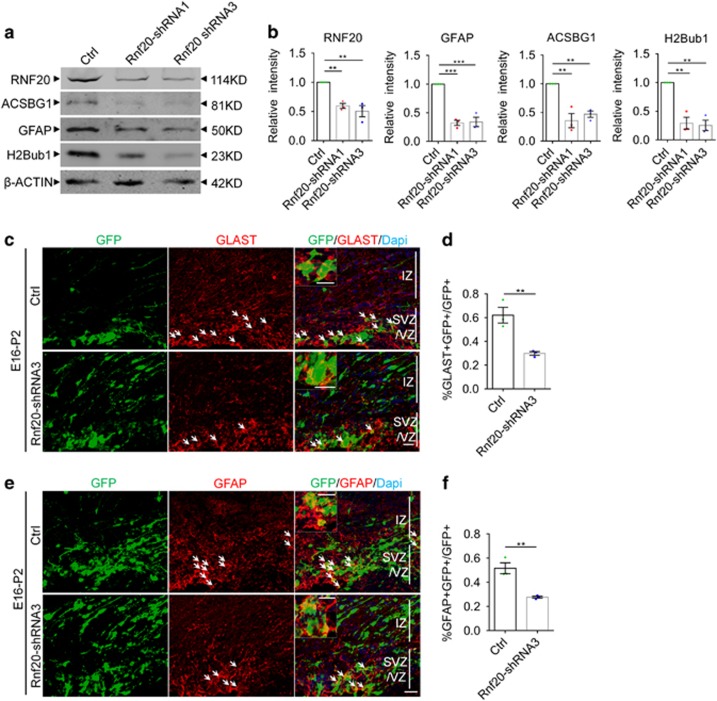
RNF20 knockdown reduces astrocyte production. (a) E16 neural precursor cells were isolated, infected and differentiated for 4 days. Western blotting showed the expression of RNF20, ACSBG1, GFAP and H2Bub1 with indicated antibodies. (b) Quantification for protein levels of RNF20, ACSBG1, GFAP and H2Bub1 in (a). Mean±S.E.M.; n=3. (c) Embryos from E16 pregnant mouse were electroporated with control (pSicoR-GFP) or Rnf20-shRNA3 plasmids. The percentage of GLAST-positive astrocytic precursor cells was investigated at P2. The arrows indicate GLAST-positive astrocytic precursor cells that were colabeled with GFP-positive cells. Enlarged images showed the colocalization of GLAST and GFP. (d) Quantification for the percentage of GLAST-positive astrocytic precursor cells in all of the GFP-positive cells in VZ, SVZ and IZ. Mean±S.E.M.; n=3. (e) E16 embryos were subjected to in utero electroporation and analyzed at P2. Immunostaining for GFAP was performed. The arrows indicate GFAP-positive astrocytes that were colabeled with GFP-positive cells. Enlarged images showed the colocalization of GFAP and GFP. (f) Quantification for the percentage of GFAP-positive astrocytes in all of the GFP-positive cells in VZ, SVZ and IZ. Mean±S.E.M.; n=3. **P<0.01, ***P<0.001, one-way ANOVA for (b), Student’s t-test for (d) and (f). Scale bar, 20 μm

