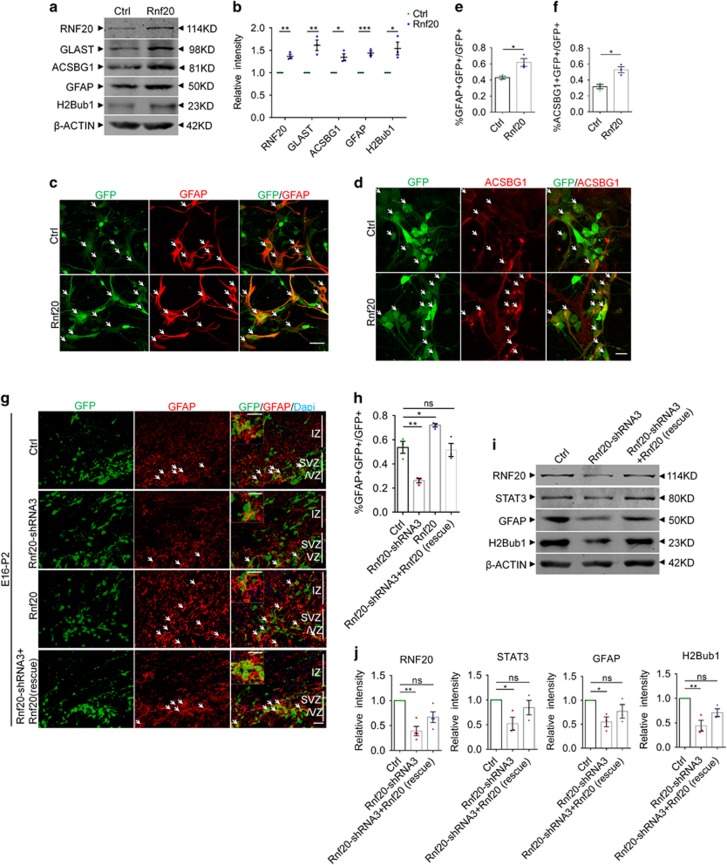
Figure 5.
RNF20 is sufficient for astrocytic differentiation. (a) E16 neural precursor cells were isolated, infected and differentiated for 4 days in vitro. Western blotting showed the expression of RNF20, GLAST, ACSBG1, GFAP and H2Bub1 with indicated antibodies. (b) Quantification for protein levels of RNF20, GLAST, ACSBG1, GFAP and H2Bub1 in (a). Mean±S.E.M.; n=3 (n=4 for RNF20). (c and d) Immunostaining showed that overexpression of RNF20 increased the production of GFAP- or ACSBG1-positive astrocytes. E16 neural precursor cells were isolated, infected and differentiated for 4 days in vitro. Scale bar in (c) 50μm, in (d) 20 μm. (e and f) The percentage of GFAP- or ACSBG1-positive cells in all the infected GFP-positive cells in (c) and (d) was quantified. Mean±S.E.M.; n=3. (g) Embryos from E16 pregnant mouse were electroporated with control, Rnf20-shRNA3, Rnf20 or co-electroporated with Rnf20-shRNA3 resistant-Rnf20 and Rnf20-shRNA3, and brains were harvested and analyzed at P2. Immunostaining for GFAP was performed. Enlarged images showed the colocalization of GFAP and GFP. Scale bar, 20 μm. (h) The percentage of GFAP-positive astrocytes in GFP-positive cells in VZ, SVZ and IZ was quantified. Mean± S.E.M.; n=3. (i) E16 neural precursor cells were infected with control or Rnf20-shRNA3 lentivirus, or co-infected with Rnf20-shRNA3 resistant-Rnf20 and Rnf20-shRNA3 lentivirus, and differentiated for 4 days in vitro. Western blotting showed the rescue experiments in vitro. Expression of RNF20, STAT3, GFAP and H2Bub1 were detected with indicated antibodies. (j) Quantification for protein levels of RNF20, STAT3, GFAP and H2Bub1 in (i). Mean±S.E.M.; n=3. *P<0.05, **P<0.01, ***P<0.001. NS, not significant; Student’s t-test for (b, e and f), one-way ANOVA for (h) and (j)

