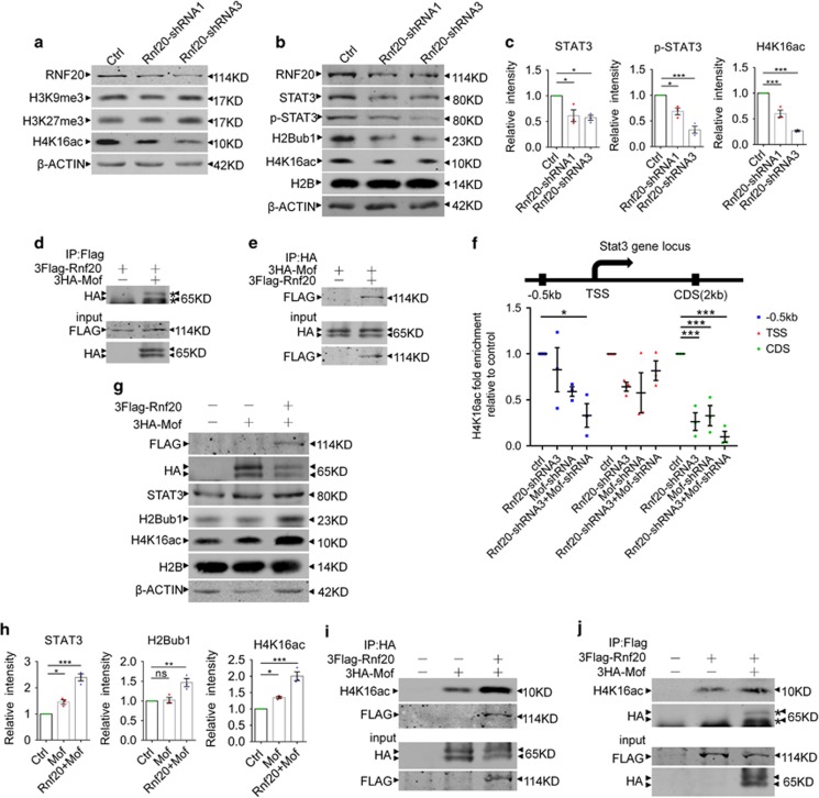
Figure 7.
RNF20 recruits MOF to Stat3 gene locus to acetylate H4K16. (a) Histone modifications screening after RNF20 knockdown via lentivirus infection in E16 primary neural precursor cells followed by 4 days differentiation. Histone modifications were detected with the indicated antibodies. (b) Western blotting showed the expression of RNF20, STAT3, p-STAT3, H2Bub1 and H4K16ac after lentivirus infection in E16 neural precursor cells followed by 4 days differentiation. (c) Protein levels of STAT3, p-STAT3 and H4K16ac in (b) were quantified. Mean±S.E.M.; n=3. (d and e) Co-IP assay was performed to examine the interaction between RNF20 and MOF. In (d), N2a cells were co-transfected with 3Flag-Rnf20 and control plasmids, or 3Flag-Rnf20 and 3HA-Mof plasmids. In (e), N2a cells were co-transfected with 3HA-Mof and control plasmids, or 3Flag-Rnf20 and 3HA-Mof plasmids. After 48–72 h, anti-Flag (d) or anti-HA (e) beads were used to pull down the proteins. Results were visualized by western blotting with the indicated antibodies. (f) Ch-IP assay showed knockdown of RNF20 promoted the reduction of H4K16ac caused by MOF knockdown at Stat3 coding region and the promoter. N2a cells were transfected with indicated plasmids and cultured for 48–72 h. (g) Western blotting showed that co-overexpression of 3Flag-Rnf20 and 3HA-Mof promotes more H4K16 acetylation and STAT3 expression than overexpression of single 3HA-Mof. N2a cells were transfected with indicated plasmids and cultured for 48–72 h. Results were visualized with indicated antibodies. (h) Protein levels of STAT3, H2Bub1 and H4K16ac in (g) were quantified. Mean±S.E.M.; n=3. (i and j) Co-IP assay showed that more H4K16ac was pulled down with anti-HA beads or anti-Flag beads in the 3Flag-Rnf20 and 3HA-Mof co-overexpressed group than in the 3HA-Mof or 3Flag-Rnf20 group. In (i and j), N2a cells were transfected with indicated plasmids and cultured for 48–72 h. Western blotting in (i) and (j) were performed with indicated antibodies. *P<0.05, **P<0.01, ***P<0.001, one-way ANOVA

