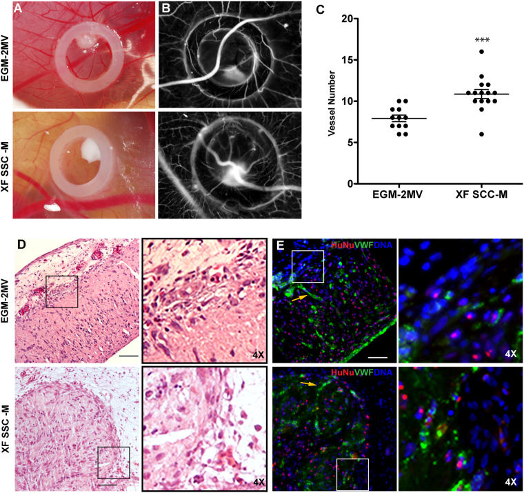
Figure 6.
In vivo CAM assay. The angiogenic potential of spheroids in both conditions was tested using chick embryonic membrane. (A) Brightfield images of spheroids inside the CAM O-ring; (B) photomicrographs acquired for counting newly formed vessels; (C) Angiogenic effect of spheroids grown in the standard versus XF conditions, expressed by number of newly formed vessels (n ≥ 12, two independent experiments, ***p < 0.0004, Mann-Whitney test was used for statistical comparison). (D) HE staining of CAM interface and spheroids after 72 h incubation time (Scale bar 100 μm). (E) Immunofluorescence of equivalent sections represented in D (Scale bar 50 μm). vWF (green) was used to access endothelial organization, HuNu (red) to discriminate human nuclei and DAPI (blue) for total nuclei. In images (D) and (E), insets (and respective 4× magnifications, depicted on the right) highlight the direct contact between spheroids and the CAM. Yellow arrows are pointing to organized OEC inside the compacted spheroids.

