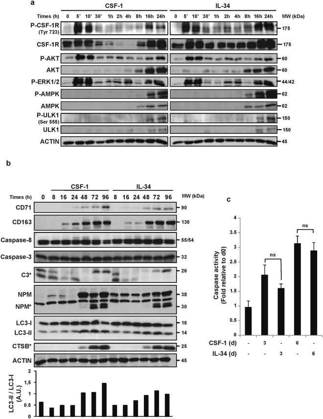
Figure 2.
Caspases and autophagy are activated upon IL-34 or CSF-1 treatment. Human peripheral blood monocytes from healthy donors were exposed to 100 ng/mL CSF-1 or 100 ng/mL IL-34 for the indicated times. (a) Immunoblot analysis of indicated proteins in monocytes following CSF-1 or IL-34 stimulation. P indicate phosphorylated proteins. Each panel is representative of at least 3 independent experiments. (b) Immunoblot analysis of indicated proteins in monocytes following CSF-1 or IL-34 stimulation. The ratio of the LC3-II protein level to that of LC3-I protein level was determined using ImageJ software. Actin was detected as the loading control. Asterisks indicate cleavage fragments. Each panel is representative of at least 3 independent experiments. (c) Caspase activity was quantified by flow cytometry analysis using DEVD-FITC. The results are expressed as the fold induction compared with untreated cells and represent the mean ± SD of 3 independent experiments performed in duplicate. n.s. denotes non-significant according to a paired student t test.

