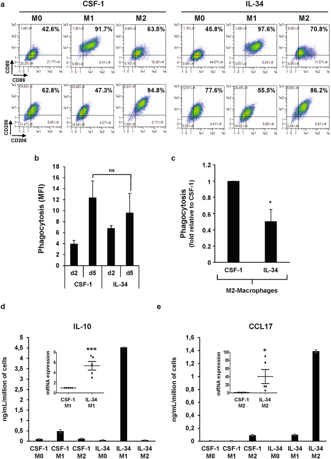
Figure 4.
IL-34 macrophages have a different polarization potential as compared to CSF-1-macrophages. (a) Human monocytes were differentiated during 7 days with 100 ng/mL CSF-1 or 100 ng/mL IL-34 and then polarized into M1-macrophages (LPS + IFNγ) or M2-macrophages (IL-4) for 2 days. Macrophage polarization was evaluated by 2-color flow cytometric analysis. (b) Functional assay of monocytes exposed for 2 or 5 days to 100 ng/mL CSF-1 or 100 ng/mL IL-34. The results are expressed as MFI and represent the mean ± SD of 3 independent experiments performed in duplicate. n.s. denotes not statistically significant according to a paired student t test. (c) Functional assay of monocytes exposed for 7 days with 100 ng/mL CSF-1 or 100 ng/mL IL-34 and then polarized into M2-macrophages (IL-4) for 2 days. The results are expressed as the fold induction compared to CSF-1 macrophages and represent the mean of 3 independent experiments performed in duplicate. *P < 0.05 according to a paired student t test (versus CSF-1-macrophages). (d,e) Human monocytes were differentiated during 7 days with 100 ng/mL CSF-1 or 100 ng/mL IL-34 and then polarized into M1-macrophages (LPS + IFNγ) or M2-macrophages (IL-4) for 24 hours. The expression of the indicated mRNA was analyzed by qPCR (mean ± SEM of 5 independent experiments). *P < 0.05, ***P < 0.001 according to a paired student t test (versus CSF-1 macrophages). The production of IL-10 and CCL17 was analyzed using Multi-Analyte ELISArray kit as described in Material and Methods section. The results are expressed as ng/mL per million of cells and represent the mean ± SD of 2 independent experiments performed in duplicate.

