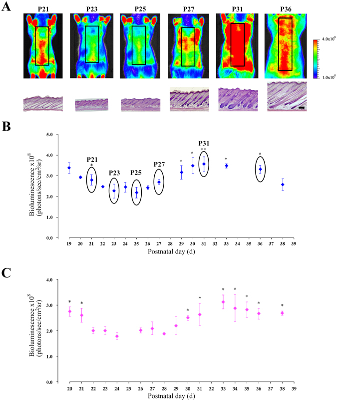
Figure 3.
In vivo bioluminescence imaging of NG2-fLuc Tg rats during the first postnatal hair cycle. (A) Bioluminescence images of male Tg rats from P19 to P38 during the first postnatal hair cycle; black squares in each image show regions of interest (ROI). Haematoxylin-stained images of skin from Tg rats visualized by bioluminescence imaging at each time point; scale bars; 500 μm. (B and C) Quantification of bioluminescence signals from ROI in the dorsal surface skin of male (N = 14, blue diamonds in B) and female (N = 8, pink diamonds in C) Tg rats during the first postnatal hair cycle. Data from each animal were presented as means ± SD. *P < 0.05 and **P < 0.01; compared with telogen phase (P23 in B or P24 in C).

