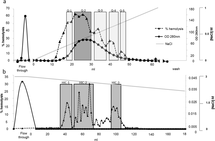
Figure 4.
Separation and partial purification of an actinoporin-like hemolysin from Stylophora. In the chromatograms, the shaded areas represent active fractions that were pooled and submitted either to re-chromatography (after anion exchange) or two tandem mass spectrometry (after hydrophobic interactions chromatography). The straight grey lines represent the gradient of salt used for chromatography. (a) Separation of Stylophora crude extract by anion exchange chromatography on a Q- sepharose FF 18.5 ml column. (b) Separation of pool Q-2 from panel A, above, on a Hydrophobic Interactions Phenyl- Sepharose HP 7.8 ml column.

