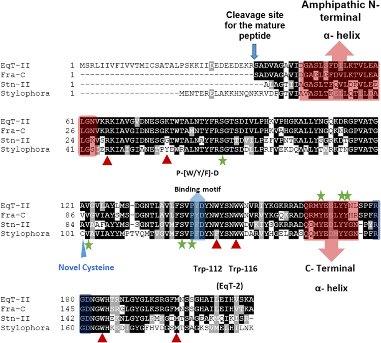
Figure 5.
Multiple alignment of the Stylophora actinoporin and three well studied sea anemone actinoporins: equinatoxin II (EqT-II, Actinia equina), stycholysin II (Stn-II, Stichodactyla helianthus) and fragaceatoxin C (Fra-C, Actinia fragacea). The regions highlighted in red are the C and N- terminal α- helixes, the red triangles point out residues that have shown to be important for membrane attachment61,69, the blue arrow shows the RGD motif involved in oligomerization105 and the green stars represent residues that have shown to be important for sphingomyelin recognition58,61. The blue region shows the conservation of the P-[W/Y/F]-D membrane binding motif106,107.

