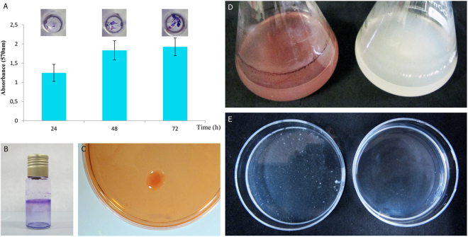
Figure 2.
Biofilms and cellulose formation. Attachment of strain PEPV40 to abiotic surfaces observed in polystyrene microtiter plates along the time (Bars indicate the standard error. The experiment was repeated three times) (A) and in borosilicate glass tubes (B). Cellulose formed by the strain in plates containing Congo Red (C) and in YMB liquid medium with Congo Red (D, left) and without (D, right). Flocs formed by the strain in YMB liquid medium (E, left) and after 2 h incubation at 37 °C in pH 5 PCA buffer containing 10 U/ml Trichoderma viride commercial cellulase (E, right).

