Figure 3.
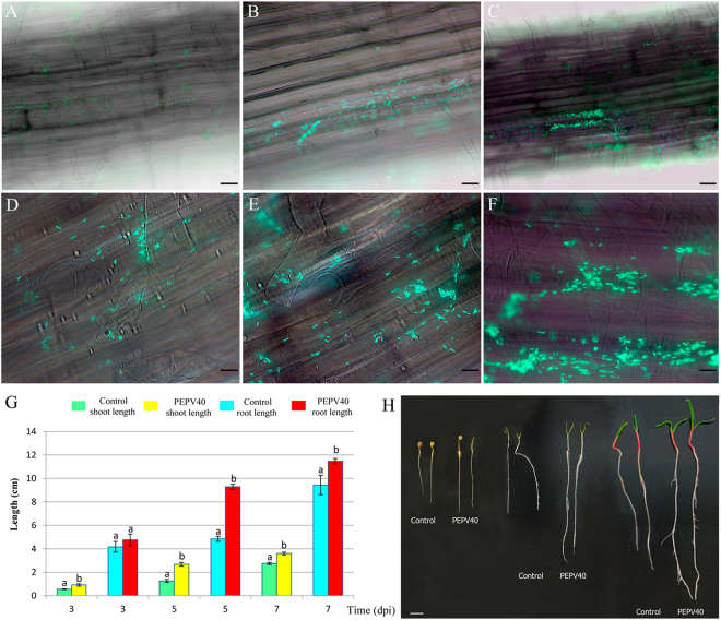
Bacterial root colonization. Fluorescence optical micrographs of spinach seedlings roots obtained at 3 (A,D), 5 (B,E) and 7 (C,F) days after inoculation with GFP-tagged cells of strain PEPV40 (A,B, and C, bar 60 µm, and D,E and F, bar 12 µm). The micrographs show the ability of strain PEPV40 to colonize the roots surfaces of spinach and the initiation of microcolonies. Effect of the strain PEPV40 inoculation in the early steps of spinach seedlings: Evolution of shoot and root length (G). Bars indicate the standard error. Histogram bars marked with the same letter in each treatment are not significantly different from each other at P = 0.05 according to Fisher’s Protected LSD (Least Significant Differences). Uninoculated and inoculated spinach seedlings from the in vitro experiment (H, bar 1 cm).

