
Figure 1.
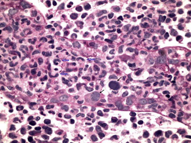
Kidney biopsy sample containing a virus-infected renal tubule. Most of the renal tubular epithelial cells are lost or damaged beyond recognition. Two of the residual epithelial cells contain “smudged” intranuclear viral inclusions typical of adenovirus, with nuclear enlargement and peripheral displacement of nuclear chromatin (black arrows). There is an associated intratubular and peritubular infiltrate consisting predominantly of distinct histiocytes with elongated, curved, and crinkled nuclei (open blue arrows). Periodic acid–Schiff stain, original magnification ×600.