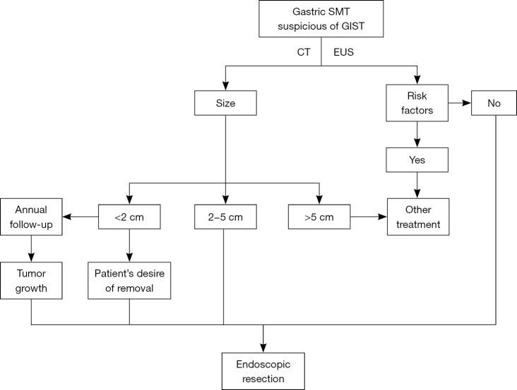
Figure 1.
The patient selection diagram of endoscopic resection for gastric GISTs in our hospital. Risk factors: ulceration or erosion at the site of tumor location; EUS shows irregular border, internal heterogeneity include anechoic area (i.e., necrosis) and echogenic loci (i.e., bleeding), heterogeneous enhancement, regional lymph node swelling; CT show metastasis or invasion out of the gastrointestinal tract; a Zubrod-ECOG performance status ≥2; have severe cardiopulmonary disease or blood coagulation disorders. GIST, gastrointestinal stromal tumor; EUS, endoscopic ultrasonography; CT, computed tomography; SMT, submucosal tumor.

