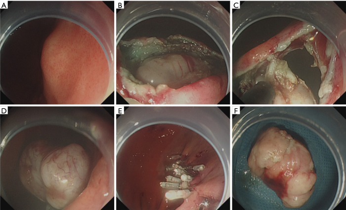
Figure 4.
Case illustration of endoscopic full-thickness resection. (A) We could see a submucosal tumor in the gastric corpus; (B) after submucosal injection, we precut and remove the mucosal and submucosal layer to expose the tumor; (C,D) endoscopic full-thickness resection of the tumor, we could see the abdominal cavity through the “active perforation”; (E) close the wound with several clips; (F) the resected tumor.

