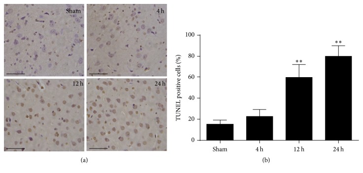
Figure 2.
Representative images of TUNEL staining of the brain cortex after ROSC. (a) TUNEL staining in the brain cortex at 4 h, 12 h, and 24 h after ROSC. Scale bar = 10 μm. (b) Quantification of the apoptotic neurons by positive staining. Data are represented as mean ± standard deviation (SD). n = 6 photomicrographs were counted per animal. ∗∗P < 0.01 versus sham.

