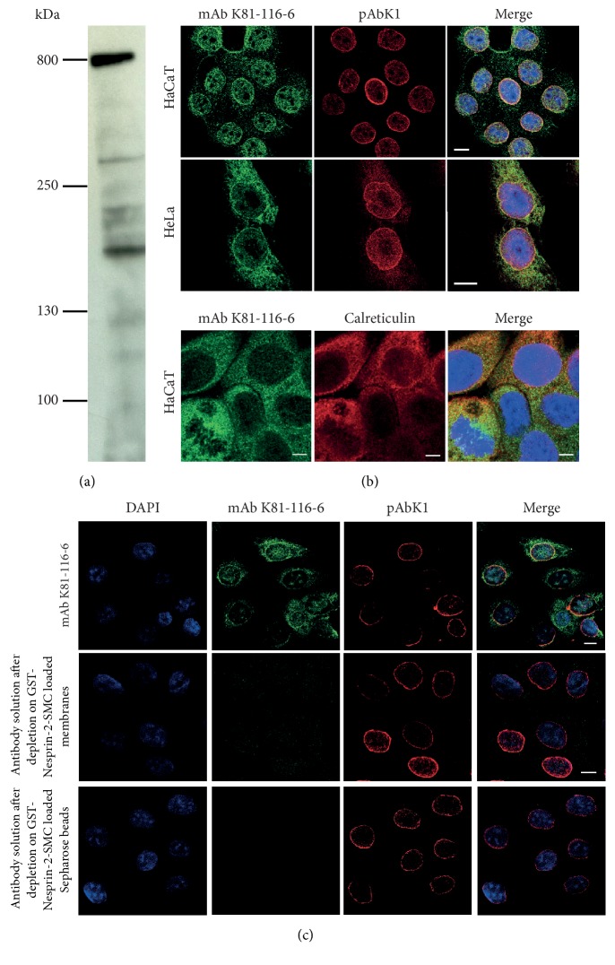
Figure 2.
Characterization of monoclonal antibodies directed against the SMC domain. (a) Detection of Nesprin-2 with mAb K81-116-6 in HaCaT cell lysates. Proteins were separated by SDS-PAGE (3–12% acrylamide). (b) mAb K81-116-6 staining of HaCaT and HeLa cells. pAbK1 was used as bona fide Nesprin-2 antibody. DAPI stains the DNA (in Merge). Bar, 10 μm. Lower panel, colocalization of Nesprin-2 detected by mAb K81-116-6 with ER marker calreticulin in HaCaT cells. Bar, 5 μm. (c) Analysis of the specificity of mAb K81-116-6. Antibodies were depleted from the hybridoma supernatant by the indicated procedures. Antibody depleted supernatants were then used for immunofluorescence analysis. Bar, 10 μm.

