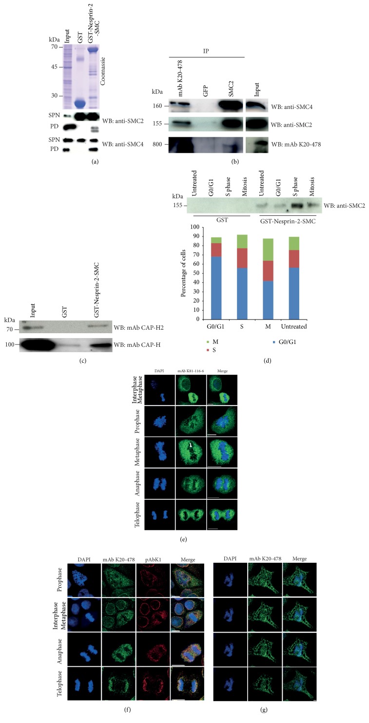
Figure 3.
Interaction of Nesprin-2-SMC and Nesprin-2 with SMC2 and SMC4. (a) Precipitation of SMC2 and SMC4 with GST-Nesprin-2-SMC from HaCaT cell lysates. Precipitates were resolved on SDS-polyacrylamide gels (10% acrylamide) and probed with SMC2 and SMC4 specific antibodies. SPN, supernatant after pulldown; PD, pulldown. The lower molecular weight band in the SMC2 pulldown is presumably a breakdown product. (b) Immunoprecipitation of SMC2 from HaCaT cell lysates with Nesprin-2 specific mAbK20-478 and of Nesprin-2 with SMC2 specific antibodies. GFP-specific monoclonal antibodies were used for control. The antibodies used for immunoprecipitation are indicated above the panels (IP). The blots were probed with the antibodies listed on the right (WB). Immunoprecipitates were resolved on gradient gels (3–12% acrylamide) and 10% acrylamide gels as appropriate. The data are from one blot; however, the input was not directly adjacent to the SMC2 IP. (c) Interaction of CAP-H2 (condensin II) and CAP-H (condensin I) with Nesprin-2-SMC. Pulldowns were performed with HaCaT cell lysates and GST for control and GST-Nesprin-2-SMC as indicated. Unsynchronized cells were used for the experiments shown in (a)–(c). (d) Analysis of the Nesprin-2-SMC interaction with SMC2 during the cell cycle. HaCaT cells were synchronized with RO-3306 or other reagents as described in Materials and Methods in order to obtain the relevant cell cycle phases. Cell cycle phases were assessed by FACS analysis; the results are depicted in the accompanying diagram. Pulldown was carried out with GST-Nesprin-2-SMC bound to GST-Sepharose. GST was used for control. The blot was probed with SMC2 specific antibodies. (e) Localization of Nesprin-2 as detected with mAb K81-116-6 (green) during mitosis in HaCaT cells. DNA was stained with DAPI. Arrow points to filamentous staining across the chromosomes. (f) Nesprin-2 distribution in HaCaT cells during mitosis as detected with mAb K20-478 (green) and pAbK1 (red). DNA was detected with DAPI. Bar, 10 μm. (g) Nesprin-2 presence on chromosomes. Different Z-stacks (from top to bottom: 0 μm, 0.21 μm 0.42 μm, and 0.84 μm) from a COS7 cell in anaphase stained with mAb K20-478. DNA was stained with DAPI. Bar, 5 μm.

