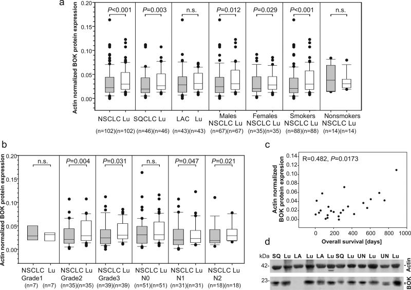
Figure 1.
BOK protein expression in NSCLC tissues and adjacent lung tissue. (a and b) Expression data are shown as median with the upper ranges of 75% (box) and 90% (whisker) and the lower ranges of 25% (box) and 10% (whisker). Statistical differences were calculated by the Wilcoxon signed rank test. P values lower than 0.05 were considered statistically significant. (c) Spearman’s correlation between BOK protein expression in lymph node positive NSCLC and patients’ overall survival. (d) A representative image of quantitative western blot of total protein lysates from NSCLC and lung parenchyma. SQ - squamous cell lung carcinoma, LA - lung adenocarcinoma, UN- undifferentiated cell lung carcinoma, Lu - lung parenchyma.

